Secrets Behind Red Blood Cells Amazing Flexibility
Oct 25, 2005 - 10:05:00 PM, Reviewed by: Dr.
|
|
�Although we made several assumptions, our model is an important step toward our goal of understanding the molecular basis of cell membrane mechanics. We think this model may explain why the deformations of red blood cells squeezing through narrow capillary openings are so important: the movement of proto-filaments may effectively enhance the diffusion of oxygen from red blood cells deep in tissues and organs where the exchange is most needed.�
|
By UCSD School of Engineering,
A human red blood cell is a dimpled ballerina, ceaselessly spinning, tumbling, bending, and squeezing through openings narrower than its width to dispense life-giving oxygen to every corner of the body. In a paper published in the October issue of Annals of Biomedical Engineering, which was made available online on Oct. 21, a team of UCSD researchers describe a mathematical model that explains how a mesh-like protein skeleton gives a healthy human red blood cell both its rubbery ability to stretch without breaking, and a potential mechanism to facilitate diffusion of oxygen across its membrane.
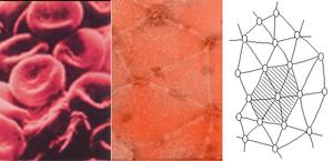
�Red cells are one of the few kinds of cells in the body with no nucleus and only a thin layer of protein skeleton under their membrane: they are living bags of hemoglobin,� said Amy Sung, a professor of bioengineering at UCSD�s Jacobs School of Engineering and coauthor of the study. �Very little is known about how the elements of the membrane skeleton behave when red blood cells deform, and we were amazed at what our simulation revealed.� Scientists have been mystified for years by the human red blood cell membrane skeleton, a network of roughly 33,000 protein hexagons that looks like a microscopic geodesic dome. Sung and her collaborators at the Jacobs School of Engineering focused on what they view is a key component at the center of each hexagon, a rod-shaped protein complex called the proto-filament. The proto-filament is 37 nanometers in length and made of a protein called actin. Elsewhere in the human body, bundles of actin form contractile muscles, and matrices of actin are responsible for the gel-like properties of various cells� cytoplasm. However, the foreshortened actin fibers in the proto-filaments act as rigid rods held in suspension by six precisely positioned fibers made of the actin-binding protein spectrin.
 |
| The human red blood cell membrane skeleton is a network of roughly 33,000 protein hexagons that looks like a microscopic geodesic dome. |
Robert Skelton, a professor of mechanical and aerospace engineering at the Jacobs School of Engineering and a co-author of the study, employed the unorthodox approach of modeling the proto-filaments as if they were part of a tensegrity structure. Artists have been more familiar with rod-and-cable tensegrity structures than scientists. Sung asked Skelton to collaborate on her red blood cell project because Skelton and his students have pioneered the development of rigorous scientific tools to analyze the movement and balance of forces in many types of tensegrity systems.
�Although we made several assumptions, our model is an important step toward our goal of understanding the molecular basis of cell membrane mechanics,� said Sung.
Sung, Skelton, and post-doctoral fellows Carlos Vera and Frederic Bossens combined mathematical modeling of a proto-filament as a tensegrity structure with a visualization technique that revealed how a single proto-filament moves in response to the pulling force of six spectrin fibers attached to it. Sung�s team was pleasantly surprised that its model also generated near-random yaw angles for the proto-filament during deformation of the red blood cell and no more than 18 degrees of pitch relative to the membrane in most cases. The modeling suggests that the more a red blood cell is mechanically deformed, the more likely its individual proto-filaments will rotate left and right like a baseball bat swung over home plate. �We think this model may explain why the deformations of red blood cells squeezing through narrow capillary openings are so important: the movement of proto-filaments may effectively enhance the diffusion of oxygen from red blood cells deep in tissues and organs where the exchange is most needed.�
 |
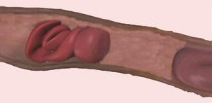
| A model proposed by researchers at UCSD's Jacobs School of Engineering may explain why the deformations of red blood cells as they squeeze through narrow capillary openings are so important. |
The team is planning to broaden its analysis to include the effects of trans-membrane proteins that physically anchor the underlying protein network to the red blood cell membrane. The team also plans to enlarge its simulation to visualize more than one proto-filament at a time, and eventually model the simultaneously movement of all 33,000 proto-filaments in a cell.

- Carlos Vera, Robert Skelton, Frederic Bossens, and Lanping Amy Sung, "3-D Nano-mechanics of an Erythrocyte Junctional Complex in Equibiaxial and Anisotropic Deformations" (2005). Annals of Biomedical Engineering. 33 (10), pp 1387�1404.
Full text of the research article
For any corrections of factual information, to contact the editors or to send
any medical news or health news press releases, use
feedback form
Top of Page
|