|
From rxpgnews.com Cytology
The process of membrane fusion is essential for the structure and dynamics of all cells in our bodies. Fusion is indispensable for intracellular vesicle traffic, which sustains the compartmental organisation of cells. Likewise, membrane fusion is the basic molecular process that controls the communication between cells via the secretion of hormones, neurotransmitters, and growth factors. Furthermore, fusion processes are also crucial for the interactions between our cells and various pathogens such as viruses and bacteria. However, in spite of the ubiquity of membrane fusion, many aspects of this process have remained rather controversial. This situation reflects the absence of well-defined protocols by which one can induce fusion in a controlled manner and subsequently study its dynamics with high temporal resolution.
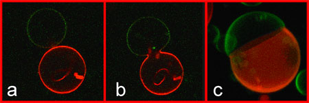
In the first protocol, artificial fusogenic molecules (a liposome whose outer wall contains molecules that cause cell fusion) or ligands, synthesized by the collaborators from Coll�ge de France, were incorporated into the lipid membranes. Two unilamellar vesicles were aspirated by two glass micropipettes. Close proximity of the vesicle membranes was achieved by displacing these micropipettes. Membrane fusion was subsequently induced by the local addition of ions that form a complex between two fusogenic molecules embedded in the opposing membranes. In the second protocol, two lipid vesicles were brought into contact by alternating electric fields. Once close contact was established, membrane fusion was induced by exposing the vesicles to an additional electric pulse. Such a pulse leads to the formation of membrane pores in the opposing membranes, which subsequently fuse in order to dispose of the edges of the pores. Both for ligand-mediated fusion and for electrofusion, the dynamics of fusion was observed using a fast digital camera with an acquisition rate of 20 000 frames per second, which corresponds to a temporal resolution of 50 microseconds. "Since previous direct imaging studies of membrane fusion were limited to time scales that exceed tens of milliseconds, the new experiments improved the temporal resolution by three orders of magnitude and revealed that the fusion process is surprisingly fast", says Rumiana Dimova, group leader in the Max Planck Institute of Colloids and Interfaces, and one of the participating scientists. Indeed, only a few hundred microseconds after the initiation of the fusion process, the fusion neck connecting the two vesicles has already reached a diameter of a couple of micrometres as shown in Figure 1(b). This implies that the fusion neck has an average expansion velocity of centimetres per second and that the initial formation of the fusion neck can be completed within about 200 nanoseconds. This is in good agreement with recent computer simulations of tension-induced fusion. In this way, the Max Planck researchers have managed to bridge the gap between theoretical predictions and available experimental knowledge about the fusion process. The experimental fusion protocols developed in the present study can be applied to other biomimetic systems and can be used to construct new ones. Particularly interesting systems, which can be studied in this way, are mixed membranes containing both lipids and fusogenic proteins such as SNAREs. One example for the construction of new biomimetic systems is provided by the formation of large vesicles with several intramembrane domains as shown in Figure 1(c). Another example consists of vesicles that contain different chemical reactants. The fusion of such vesicles initiates the corresponding chemical reactions in these rather small compartments and might be useful in order to synthesize new nanomaterials. In general, controlled membrane fusion has many potential applications in bioengineering, pharmacology, and medicine. All rights reserved by www.rxpgnews.com |
